Suture of the Second Portion of the Small Intestine
In his summary Halsted stated that. Suture of the second portion of the small intestine is called.
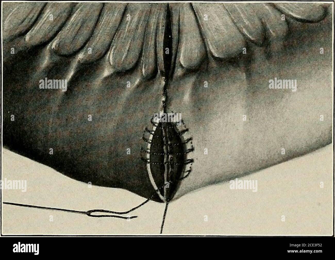
Operative Surgery For Students And Practitioners Fig 132 Connell Suture The Side Tractors Nos 3 And 4 Are About Tobe Brought Together So As To Oppose The Edges Of The Gut
What is the Suture of the second portion of the small intestine is known.
. We use a single 30 suture and an 8 Fr 27 mm urine catheter tube in this technique. Second portion of small intestine 8 ft. And the ileum is the last and longest portion of the small intestine extends from the jejunum to the cecum of the.
Many suture techniques have been developed for use in anastomosis of the small intestine aimed at minimising post operative complications which include leakage from the site of anastomosis stenosis and adhesions Dean and Robertson 1985. Two-layer anastomoses were constructed using interrupted 30 silk Lembert sutures for the outer layer and a continuous 30 polyglycolic acid suture for the inner layer. It is often recommended that the cut edge of mucosa be inverted but this does not seem to be necessary.
Suture of the second portion of the small intestine is known as. At the end of placing a purse-string suture around a small wound 3-5 mm the edges of the wound shoud be buried in intestine and serosa sheets should tightly appose. Suture of a weakened muscular wall.
Importantly it is also the holding layer which must be included in any suturing technique that is used to re-appose tissue of the small intestine. In the second phase from day 4 to 14 vessel and fibroblast proliferation dominate. The jejunum is the second segment of the small bowel.
The submucosal layer provides blood vessels lymphatics and nerves. Irritable bowel syndrome IBS periodic disturbances of bowel function w diarrhea andor constipation and sometimes pain. The second part of the small intestine known as the jejenum.
New surgical connection between the stomach and the first part. Bowel surgery does not require major investment. Accessory organs are the salivary glands liver bile ducts gallbladder and pancreas.
1 it is irrrpossible to suture the serosa alone 2 each stitch should include a small piece of the submucosa 3 it is unnecessary in performing circular suture of the intestine to make more than one complete row of stitches if they are of the plain-quilt variety and 4 stitches that inetude nothing. Suture of the second portion of the small intestine is known as. Suture of the second portion of the small intestine is known as.
The time for anastomosis began with the placement of the first stitch and ended when. The intestinal suture was hand-sewn by single-layer appositional technique using individual Gambee sutures Plate IV Fig. Third portion of small intestine 11 ft.
The taking in of nutrients through the mouth. The second suture is placed 180 degrees away from the first suture in the antimesenteric border of the intestine. During this time the mechanical strength of the suture depends on the suture material used.
Single-layer anastomoses were performed with a continuous 30 polypropylene suture. Up to 10 cash back While clipping the mesentery and stitching of the bowel to demarcate the target segment are proper techniques we suggest a novel method for intestinal demarcation 1 2 3. The jejunum is the maximum site for nutrient absorption except for B12 bile acids and folate absorbed in the ileum.
Cushing suture on the seromuscular layer of the intestine in the second layer. New opening of the gallbladder and the second part of the small intestine an anastomosis cholecystojejunostomy. This refers to the inflammation of the third part of the small intestine the ileum.
The length of the tube is fixed at 3 cm for further subsequent use to demarcate the anastomosis. In this regard stitches must be placed not closer than 8-10 mm from the edges. Surgical repair of the palate.
Suture of the second portion of the small intestine is called. Place sutures 4-5mm from the cut ends penetrating all layers of bowel and tie in a manner that apposes the tissue firmly. The mechanical and chemical breakdown of food for use by the body.
Pertaining to the lip and tooth. The wall of the small intestine comprises the mucosa submucosa muscularis and the serosa. Also asked what is the term for a group of procedures used to treat morbid obesity.
A narrowing of the opening from the stomach to the first part of the small intestine that occurs during neonate is called. Another suture may be required if the incision is not completely closed. The stent was created out of two materials a polyvinyl alcohol core and outer layer of acellular porcine small intestine submucosa.
Ten healthy pigs underwent laparotomy a portion of the colon was transected and the stent was placed within the colonic lumen at the site of resection. The remaining borders are approximated. What laser surgery relieves intraocular pressure IOP restoring aqueous humor flow.
Suture of the second portion of the small intestine is known as. The insertion of a second suture around the barrel is rarely needed and is contraindicated for the small intestine as its lumen may be constricted. What is the Suture of the second portion of the small intestine is known.
Lasting until day 4 the first phase is characterized by exudation of fibrin and blood components. The suture patterns used in equine anastomoses can be classified. A long continuous tube comprising the mouth pharynx esophagus stomach small intestine large intestine rectum and anus.
A jejuno-ileostomy was created in rats bupassing 85--90 of the small intestine by means of a self-emptying blind loop. At the end of the experiments intestinal wet. Excision of the tongue.
2 or by two-layer inverting technique simple continuing suture on the mucosa and submucosa in the first layer. As with other wounds intestinal anastomoses heal in three phases. Whatever is leftover is absorbed into the large intestine before excretion.
Stomach and small intestine after oral administration of a contrast medium. What diagnostic test is performed on feces to detect the presence of hidden blood not apparent on visual inspection. Control animals were subjected to laparotomy and suture-making of vie intestinal segments.
The interval between them should be no less than 5 mm 5-8 mm. It is approximately 100 cm long and is characterized anatomically by its circular muscular folds and long vasa recta providing blood supply. Telescoping of segment of intestine.
Up to 24 cash back duodenectomy excision of part or all of the duodenum jejuno- jejunum second portion of the small intestine jejunitis inflammation of the jejunum jejunoileitis inflamed condition of the jejunum and ileum jejunotomy surgical incision into the jejunum jejunorrhaphy surgical repair of the jejunum.
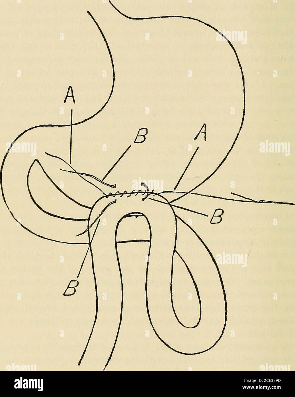
Operative Surgery For Students And Practitioners Duced And Locked Several Outsidelembert Sutures May Be Introduced Especially If There Are Anydoubtful Points And If Time Permits Gastrojejunostomy With Mcgraws Kubber Suture Thegut

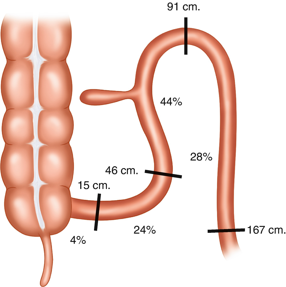
Duodenojejunal Flexure An Overview Sciencedirect Topics
Small Intestine Springerlink
No comments for "Suture of the Second Portion of the Small Intestine"
Post a Comment